|

|
|
|
|
|
|
|
|
|
|
|
|
|
|
|
Obermedizinalrat Dr. med. univ. Ferdinand
SILBERBAUER
|
|
|
Radiosensitizing
by heavy atoms –
Approach to a
new more efficient radiation therapy
keywords: Radiosensitizing by
heavy atoms; enhancement of focal dose by heavy atoms
Summary:
In this
report the beginning of a new, more effective kind of high
voltage therapy is shown, while as much heavy atoms as
possible are selectively brought into a malignoma, which
liberate much more energy in the tumour, as it otherwise may
be the case. A great part of the radiation energy, what
would simply penetrate the tumor without damaging it, is
forced by the heavy atoms to get free selectively in the
tumor. As more electrons, according to the ordinal number of
the heavy atoms, are existing in the coat of an atom, and as
more of these heavy atoms are included in a tumor, as more
additional energy is liberated in the tumor in the high
voltage therapy. This circumstance leads to healings, where
they wouldn't be possible in other case, and promises for
the future to need only half of the now usual radiation dose
and to achieve healings with an on certainty bordering
probability.
Introduction
In this
report will be explained, why heavy atoms, which are
incorporated in a tumour, induce an enhanced focal dose in
the radiation therapy: by the enrichment of heavy atoms in a
tumor (iodine, gold, gadolinium or platinum) is caused here
during a high voltage irradiation an enhanced formation of
positrons and electrons. Conditioned through that, by the
mutual deletion of positron and electron the so-called
extermination - radiation with the energy of 0,511 MeV is
created. This extermination - radiation causes now in the
tumor the Compton - photo - complex - reactions, which take
place in a very small area (within millimeters) closely
related to the heavy atoms, i.e., at an high - energy –
Compton - effect with an heavy atom a shower of electrons,
which damages the tumour, is released, and the resulting low
energetic Compton - scattered - radiation is absorbed by a
light atom ( f.i. oxygen, carbon, nitrogen) within a mean
distance of 2mm off the heavy atom by means of a photo-
effect,
what also damages the tumour. In addition the middle
energetic Compton - scattered - radiation (around
100 KeV) from the surrounding tissue is absorbed by
photo- effects with high incidence proportional to the fourth
potency of the effective ordinal number of the tumour in the
heavy - atom - loaded tumour tissue. Moderately heavy atoms,
like iron or zinc, if they are accumulated in the tumour,
they induce an increased photo - absorption of the middle
and low energetic Compton - scattered - radiation from the
tissue surrounding the tumour.
Now in the
high voltage therapy the effects of the heavy atoms in a
tumour are first theoretically and then by concrete
instances discussed. Already some tens of years ago in the
radiation physics [1, 2] the basis of this new start of
radiation therapy has been explored, what has the aim to
incorporate as much as possible heavy atoms selectively into
a malignoma. Heavy atoms have the ability to take much more
energy out of high voltage photons by high energetic Compton
effects [1, 2], as what it is possible by light atoms. In
this case many electrons are released all at once out of a
heavy atom, by what a considerable part of energy of a high
voltage photon is consumed [1, 2]. These released electrons
release yet secondary and tertiary electrons [2] and disturb
and change the biochemical bindings of the chromosomes, by
what the death of the cells is caused. The remaining Compton
- scattered - photons are mainly absorbed by means of photo
- absorption [1, 2], by what also electrons are released,
which operate destroying in the tumour. Worthy readers,
please save me from quoting mathematical formulas for the
expected effects! I found 1989 [6] mathematical nearings,
which showed, that the probability of electron - pair -
formation [1, 2], Compton effects and photoelectric effects
in the case of incorporation of heavy atoms into a tumour is
increased and that the enhancement, which is achieved in the
CT, can be employed for the calculation of the expected
focal dose [6].But it is difficult to introduce energetic
levels from 1 MeV up to 25 MeV stepwise into a mathematical
formula. Just so difficult is the introduction of secondary
and tertiary electrons into this formula. Also the ordinal
number of the employed heavy atoms plays a bigger role as
expected [3,4,10,11,12,13]. In the future we should let us
guide by empiricism. In the reality the effects seem to be
much stronger than these by me at that time calculated [
3,4,5,8,10,11,12,13].
When
during high energetic Compton - effects low energetic
Compton - scattered - photons are released and within a mean
radius of two millimetres are absorbed again, so it is
possible to call these proceedings as a Compton - photo -
complex - reaction [6].
By these
Compton - photo - complex - reactions in the low voltage
range 25 years ago a nine year old boy, who was suffering
from an inoperable astrocytoma IV in the brain stem, was
cured. By erosion haemorrhages an accumulation of iron
pigment had occurred in the 20 millimetres measuring tumour,
what was visible in the CT. The radiation therapy with
cobalt 60, caused by me, leaded with a focal dose of only
50Gy to a complete deletion of the tumour. Ten years after
the radiation therapy the young man was completely
rehabilitated. Similar effects every time could be obtained
by contrast medium assisted radiation therapy, contrast
medium, as it is used at the CCT, to cure patients with an
astrocytoma, meningeoma or neurinoma or metastases of
malignomas in the brain [10,11,12,13]. These tumours namely show at the CCT an
enhancement till 20 Hounsfield - units, what is caused by
the iodine atoms.
Imagine,
from astrocytoma IV suffering absolute death - candidates
could be cured already tomorrow by administering the
contrast medium 30 to 60 minutes before the irradiation with
a contrast medium - dose like at the CCT and in that time -
window, in which the enhancement is already weakly visible
[10,11,12,13]. Focal dose: 20 to 30 Gy in 4 to 6 fractions,
each 5 Gy [10].
This
assertion is confirmed by tasks from USA [3, 4] with a great
number of patients, which tell about radiosensitizing by
bromodeoxyuridine (BDU) and jododeoxyuridine (JDU). Iodine in
the JDU is distinct heavier than bromine in the BDU. Iodine
has in its atom - coat 53 electrons, bromine only 35 [1].
Iodine therefore can deliver more electrons, when a high -
voltage – photon "strikes" [1]. Although JDU and BDU
biochemically behave identically, it comes in the
astrocytoma IV with JDU as a radiosensitizer to a two - year
- surviving - quota of 68%, with BDU only of 28%. But it
must be considered, that with these substances an
enhancement at the CT not yet is discernable. Therefore a
focal dose of 60Gy is needed still for these effects. In the
Nose - Ear and Throat - field there was Cisplatinum used as
a radiosensitizer for advanced head and neck - cancers [5]
and a two year - surviving - quota of 53% and a five - year
- surviving - quota of 32% achieved. Platinum has 78
electrons in its atom - coat and leads therefore to these
astonishing results.
But with an iodine - caused just yet visible enhancement (time
window!) of an astrocytoma IV you will come out with a distinct
smaller focal dose, while a complete deletion of the tumour is
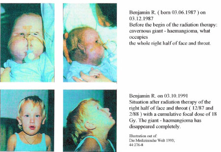
obtained [10, 11, 12, 13]. Professor Pfab in Marburg on the Lahn
in Germany achieved at a cavernous gigantic haemangioma [8] in
the head - and - neck - field by a focal dose of only 18 Gy with
hard photons of 10 MeV a complete disappearance of the tumour.
In Lissabon were cavernous haemangiomas of the liver stopped
with telecobalt by a focal dose of only 20 to 30 Gy [9]. These
results were achieved by iron, which exists as middle - heavy
atom with the ordinal number 26 abundant in the red blood cells
of the streaming blood by high energetic Compton - effects. The
electrons damage the vascular wall, before they are captured
again by the iron - atoms, what guides to disappearance of the
haemangioma or at least to a stop of its growth.
You see, a
higher energy level of the photons leads earlier to a
disappearance of the tumour, when heavy atoms are used as a
radiosensitizer, as heavier the atoms, as better the results. In
the last two years were by Gadolinium - Tex, a new magnetic
contrast medium and radiosensitizer [11,12,13], numerous various
cancers by MRI explored and then by a focal dose of only 30 Gy
insolated. In the occurred excellent results has gadolinium with
the ordinal number 64 decisive participated.
The radiation
therapy is here performed in a time - window between two and
four hours after the intravenous injection, because the
malignoma is here selectively enriched with gadolinium. In this
case the tumour is deleted without considerable damage of the
normal tissue [12, 13]. The results could be considerable
improved, if a linear accelerator of 6 to 10 MeV would be used
[8, 9].It would be possible to come out with a focal dose of 15
Gy, only the fourth part of the now usual focal dose, which
deletes yet the tumour.
At the
radiation therapy in the head - and - neck - field the following
problem occurs: when the focal dose of the high voltage therapy
becomes bigger than 30Gy, so arises with increasing dose a
painful mucositis [7] and gradual also an atrophy of the
salivary glands. Similar results you find in other fields, where
radiation therapy is used.
C
o n c l u s i o n
In the future
it will be necessary to use such a successful radiosensitizer,
which obtains with a focal dose of only 30 Gy the secure deletion of the
tumour cells. This demand I want to extend on all
tumours, which are treated by radiation therapy.
I think that
it will come to progresses in this direction by this report.
Thanks
I thank Mrs.
Dr. Brigitte Kaik for helps in English language.
Also Mrs.
Webernig Sabine, Webernig Alexander - Ingo and Mr. Waldbauer
Guenther for helps in manuscript preparation.
Many thanks to
Mr. Schlögl Reinhold and all friends, who send me very good
literature.
R e f e r e n c e s
1.
Gerthsen-Kneser-Vogel, PHYSIK, Springer Verlag
2. Theodor Laubenberger,
Leitfaden der medizinischen Röntgentechnik, Diagnostik,
Dosimetrie, Strahlenschutz, Strahlentherapie; Deutscher
Ärzteverlag
3. C.
Urtasun, D. Cosmatos, J. DelRowe, T.J Kinsella, S. Lester,
T. Wasserman, and D.S. Fulton; Jododeoxyuridine (IUdR)
combined with radiation in the treatment of malignant glioma:
a comparison of short versus long intravenous dose schedules
(RTOG 86-12)
4. H.S.
Greenberg, W.F. Chandler, W.D. Emsminger, H. Sandler,
L.Junck, M.A. Page, D.Crane, P. Mc Keever, R.Tankanow and J.
Bromberg; Radiosensitisation with carotid intra-arterial
bromodeoxyuridine + 5-fluoruracil biomodulation for
malignant gliomas; NEUROLOGY, September 1994 441715
5.
Prakash B. Chougule, Steve Suk, Quyen D. Chu, Louis Leone,
Peter T. Nigri, Robert McRea, Mary Lekas, Anthony Barone,
Dinesh Bhat and Josef Bellino; Cisplatin as a Radiation
Sensitizer in the Treartment of Advanced Head and Neck
Cancers.; CANCER, Vol. 74, No 7, October 1, 1994
6.
F. Silberbauer; Strahlentherapie mit erhöhter Wirksamkeit,
RADIOLOGE (1989) 29:48-49
7. Doz. Dr. M. Grasl,
Univ.Klinik f. HNO-Krankheiten Wien;
Schleimhaut-Irritationen bei Bestrahlung und Chemotherapie
im HNO-Bereich; Published only, 1995, Vienna
8. H. Proske, R.Pfab;
Indikationsstellung zur Hochvoltbestrahlung kavernöser
Riesenhämangiome, eine eindrucksvolle Kasuistik; DIE
MEDIZINISCHE WELT 1993; 44:276-8
9. L.Gaspar, F.
Mascarenhas, M. Sa da Costa, J. Schaller Dias, J. Gamma
Alfonso, M.E. Silvestre; Radiation therapy in the
unresectable cavernous hemangioma of the liver; RADIOTHERAPY
AND ONCOLOGY 29 (1993) 45-50
10. K.S.
Iwamoto, A. Norman, D.B. Freshwater, M. Ingram and R.G.
Skillan; Diagnosis and treatment of spontaneous canine brain
tumours with a CT scanner; RADIOTHERAPY AND ONCOLOGY 26
(1993) 76-78
11. Stuart
W. Young, Fan Qing, Anthony Harryman, Jonathan L. Sessler,
William C. Dow, Tarak D. Mody, Gregory W. Hemmy, Yunpeng Hao,
and Richard H. Miller,; Gadolinium (III) Texaphyrin: A
tumour selective radiation sensitizer that is detectable by
MRI; Proc.Natl.Acad. Sci. USA, Vol. 93, PP. 6610-6615, June
1996, Medical Sciences
12. DI
Rosenthal, C. Becerra, E. Frenkel, P. Nurenberg, D. Carbone,
S. Young, R. Miller, J. Engel, M. Holm, M.F. Renschler;
Phase I single dose trial of the radiation sensitizer
Gadolinium-Texaphyrin confirms tumour selectivity; Published
Only, AACR/ASCO Meeting 1997
13. P.
Carde, DI Rosenthal, C. Koprowsky, R. Schea, J. Ruckle, R.
Tishler, S. Qoung, R. Miller, M. Holm, M.F. Renschler; A
Phase IB/II multi-dose trial of gadoliniumtexaphyrin as a
radiosensitizer in patients with brain metastases:
preliminary results.; Tuesday May 20, 1997, Poster
discussion Session: Head and Neck and Central Nervous System
Tumours ( A 108-112) AACR/ASCO Meeting 1997
back
|
|
|
|
|
|
|
|
|
